Mandible X Ray Anatomy . The mandible is comprised of a body and paired rami, coronoid processes, and condylar processes. The mandible is the single midline bone of the lower jaw. It consists of a curved, horizontal portion, the body, and two. The mandible is the largest bone of the fascial skeleton (viscerocranium). The midline of the body is the mandibular symphysis (fig. No fracture or dislocation is seen. Radiography represents the first level imaging technique in patients with traumatic injury of the mandible. Follow the line of the mandible. Besides the bones of the middle ear, the mandible is the only mobile bone in the skull. At birth, the mandible consists of two lateral halves united in the midline at the symphysis by a bar of cartilage (fig. The ramus meets the body at the angle.
from www.wikiradiography.net
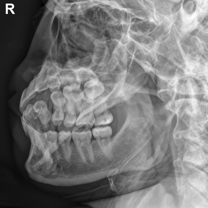
Besides the bones of the middle ear, the mandible is the only mobile bone in the skull. The mandible is comprised of a body and paired rami, coronoid processes, and condylar processes. The midline of the body is the mandibular symphysis (fig. The ramus meets the body at the angle. Radiography represents the first level imaging technique in patients with traumatic injury of the mandible. The mandible is the single midline bone of the lower jaw. Follow the line of the mandible. It consists of a curved, horizontal portion, the body, and two. At birth, the mandible consists of two lateral halves united in the midline at the symphysis by a bar of cartilage (fig. The mandible is the largest bone of the fascial skeleton (viscerocranium).
Imaging Mandibular Fractures wikiRadiography
Mandible X Ray Anatomy The midline of the body is the mandibular symphysis (fig. The mandible is comprised of a body and paired rami, coronoid processes, and condylar processes. The midline of the body is the mandibular symphysis (fig. The ramus meets the body at the angle. At birth, the mandible consists of two lateral halves united in the midline at the symphysis by a bar of cartilage (fig. The mandible is the single midline bone of the lower jaw. Radiography represents the first level imaging technique in patients with traumatic injury of the mandible. The mandible is the largest bone of the fascial skeleton (viscerocranium). It consists of a curved, horizontal portion, the body, and two. Follow the line of the mandible. Besides the bones of the middle ear, the mandible is the only mobile bone in the skull. No fracture or dislocation is seen.
From mungfali.com
Mandible Mandible X Ray Anatomy Follow the line of the mandible. The mandible is the largest bone of the fascial skeleton (viscerocranium). It consists of a curved, horizontal portion, the body, and two. The ramus meets the body at the angle. The mandible is the single midline bone of the lower jaw. Radiography represents the first level imaging technique in patients with traumatic injury of. Mandible X Ray Anatomy.
From www.researchgate.net
Landmarks of mandible bone (top image) and panoramic xrays (a Mandible X Ray Anatomy Follow the line of the mandible. The midline of the body is the mandibular symphysis (fig. Besides the bones of the middle ear, the mandible is the only mobile bone in the skull. No fracture or dislocation is seen. The ramus meets the body at the angle. The mandible is comprised of a body and paired rami, coronoid processes, and. Mandible X Ray Anatomy.
From www.pinterest.com
Facial xray... Radiology, Facial bones, Diagnostic imaging Mandible X Ray Anatomy The mandible is comprised of a body and paired rami, coronoid processes, and condylar processes. Follow the line of the mandible. Besides the bones of the middle ear, the mandible is the only mobile bone in the skull. It consists of a curved, horizontal portion, the body, and two. The midline of the body is the mandibular symphysis (fig. Radiography. Mandible X Ray Anatomy.
From mydiagram.online
[DIAGRAM] Base Of Mandible Diagram Mandible X Ray Anatomy Follow the line of the mandible. The ramus meets the body at the angle. At birth, the mandible consists of two lateral halves united in the midline at the symphysis by a bar of cartilage (fig. It consists of a curved, horizontal portion, the body, and two. No fracture or dislocation is seen. Radiography represents the first level imaging technique. Mandible X Ray Anatomy.
From radiopaedia.org
Image Mandible X Ray Anatomy At birth, the mandible consists of two lateral halves united in the midline at the symphysis by a bar of cartilage (fig. Besides the bones of the middle ear, the mandible is the only mobile bone in the skull. It consists of a curved, horizontal portion, the body, and two. Radiography represents the first level imaging technique in patients with. Mandible X Ray Anatomy.
From www.pinterest.com
soft tissue anatomy in mandible view Dental Hygiene Student, Registered Mandible X Ray Anatomy It consists of a curved, horizontal portion, the body, and two. The mandible is comprised of a body and paired rami, coronoid processes, and condylar processes. The mandible is the single midline bone of the lower jaw. The ramus meets the body at the angle. Besides the bones of the middle ear, the mandible is the only mobile bone in. Mandible X Ray Anatomy.
From www.researchgate.net
A, Posteroanterior (PA) mandible preoperative xray showing bilateral Mandible X Ray Anatomy It consists of a curved, horizontal portion, the body, and two. The ramus meets the body at the angle. Follow the line of the mandible. At birth, the mandible consists of two lateral halves united in the midline at the symphysis by a bar of cartilage (fig. The mandible is the single midline bone of the lower jaw. Radiography represents. Mandible X Ray Anatomy.
From www.pinterest.ca
Mandible. Posterior view Basic anatomy and physiology, Medical Mandible X Ray Anatomy Radiography represents the first level imaging technique in patients with traumatic injury of the mandible. The mandible is the single midline bone of the lower jaw. It consists of a curved, horizontal portion, the body, and two. No fracture or dislocation is seen. The ramus meets the body at the angle. Follow the line of the mandible. The mandible is. Mandible X Ray Anatomy.
From anatomystructure.blogspot.com
Mandible Anatomy Radiology ANATOMY STRUCTURE Mandible X Ray Anatomy Follow the line of the mandible. The mandible is the largest bone of the fascial skeleton (viscerocranium). No fracture or dislocation is seen. The ramus meets the body at the angle. The mandible is the single midline bone of the lower jaw. The midline of the body is the mandibular symphysis (fig. The mandible is comprised of a body and. Mandible X Ray Anatomy.
From radiologic-technology.blogspot.com
Technology and Techniques in Radiology Mandible Radiographic Anatomy Mandible X Ray Anatomy The mandible is comprised of a body and paired rami, coronoid processes, and condylar processes. It consists of a curved, horizontal portion, the body, and two. The midline of the body is the mandibular symphysis (fig. Radiography represents the first level imaging technique in patients with traumatic injury of the mandible. At birth, the mandible consists of two lateral halves. Mandible X Ray Anatomy.
From ar.inspiredpencil.com
Mandible Fracture X Ray Mandible X Ray Anatomy The midline of the body is the mandibular symphysis (fig. The mandible is comprised of a body and paired rami, coronoid processes, and condylar processes. Radiography represents the first level imaging technique in patients with traumatic injury of the mandible. Follow the line of the mandible. The ramus meets the body at the angle. At birth, the mandible consists of. Mandible X Ray Anatomy.
From www.pinterest.com
Mandible, anterolateral superior view. Anatomy Pinterest Anatomy Mandible X Ray Anatomy It consists of a curved, horizontal portion, the body, and two. Radiography represents the first level imaging technique in patients with traumatic injury of the mandible. The mandible is the single midline bone of the lower jaw. The mandible is comprised of a body and paired rami, coronoid processes, and condylar processes. At birth, the mandible consists of two lateral. Mandible X Ray Anatomy.
From stockcake.com
Free Xray Vision Revealed Image Download at StockCake Mandible X Ray Anatomy The mandible is comprised of a body and paired rami, coronoid processes, and condylar processes. Radiography represents the first level imaging technique in patients with traumatic injury of the mandible. The mandible is the largest bone of the fascial skeleton (viscerocranium). Besides the bones of the middle ear, the mandible is the only mobile bone in the skull. It consists. Mandible X Ray Anatomy.
From examquiz.netlify.app
Mandible x ray position examquiz Mandible X Ray Anatomy The midline of the body is the mandibular symphysis (fig. Besides the bones of the middle ear, the mandible is the only mobile bone in the skull. The ramus meets the body at the angle. Follow the line of the mandible. At birth, the mandible consists of two lateral halves united in the midline at the symphysis by a bar. Mandible X Ray Anatomy.
From www.wikiradiography.net
Imaging Mandibular Fractures wikiRadiography Mandible X Ray Anatomy Follow the line of the mandible. Radiography represents the first level imaging technique in patients with traumatic injury of the mandible. The mandible is the single midline bone of the lower jaw. The mandible is the largest bone of the fascial skeleton (viscerocranium). It consists of a curved, horizontal portion, the body, and two. Besides the bones of the middle. Mandible X Ray Anatomy.
From suhaasdental.com
Mandible Anatomy Suhaas Dental Studio Mandible X Ray Anatomy Besides the bones of the middle ear, the mandible is the only mobile bone in the skull. The midline of the body is the mandibular symphysis (fig. The mandible is comprised of a body and paired rami, coronoid processes, and condylar processes. At birth, the mandible consists of two lateral halves united in the midline at the symphysis by a. Mandible X Ray Anatomy.
From dontforgetthebubbles.com
Mandible xrays Mandible X Ray Anatomy The mandible is the largest bone of the fascial skeleton (viscerocranium). The midline of the body is the mandibular symphysis (fig. Follow the line of the mandible. No fracture or dislocation is seen. The mandible is the single midline bone of the lower jaw. Radiography represents the first level imaging technique in patients with traumatic injury of the mandible. At. Mandible X Ray Anatomy.
From www.wikiradiography.net
Imaging Mandibular Fractures wikiRadiography Mandible X Ray Anatomy The mandible is the largest bone of the fascial skeleton (viscerocranium). At birth, the mandible consists of two lateral halves united in the midline at the symphysis by a bar of cartilage (fig. The mandible is comprised of a body and paired rami, coronoid processes, and condylar processes. The midline of the body is the mandibular symphysis (fig. It consists. Mandible X Ray Anatomy.